


Mannheimia haemolytica and Pasteurella multocida are frequently responsible for bronchopneumonia in young, growing goats and sheep, with clinical signs ranging from mild depression and respiratory disease to acute death. Treatment relies on prompt and effective antimicrobial administration and supportive care; prevention relies on proper management and husbandry.
Bronchopneumonia due to Pasteurella multocida or Mannheimia haemolytica has a cranioventral lung distribution and affects sheep and goats of all ages worldwide. Young animals around the time of weaning are affected particularly severely. It is a common cause of morbidity and death in lambs and kids, especially in those that have not received adequate colostrum or in which passive immunity is waning. The disease appears to occur most often in animals that have undergone recent stress, such as transportation, weaning, change of diet, or commingling with animals from unrelated farms. Bibersteinia (formerly Pasteurella) trehalosi causes septicemia and bronchopneumonia in lambs and has also been reported as a causative agent of bronchopneumonia in calves and goats. (Also see Pasteurellosis of Sheep and Goats.)
Etiology of Bacterial Bronchopneumonia in Sheep and Goats
Both P multocida and M haemolytica are commensals of the upper respiratory tract that can cause pneumonia either alone or in conjunction with other organisms. Primary infections with respiratory pathogens such as parainfluenza type 3 (PI-3), adenovirus, respiratory syncytial virus, Bordetella parapertussis, or, in particular, Mycoplasma ovipneumoniae appear to predispose the animal to secondary infection with Pasteurella and Mannheimia.
Pathogenesis of Bacterial Bronchopneumonia in Sheep and Goats
Stress appears to be an important factor that enables Pasteurella, Mannheimia, Mycoplasma spp, other bacteria, and viruses to multiply and impair the normal physical defense mechanisms, facilitating the invasion of lung tissue and the development of pneumonia. In calves, alveolar macrophage function is impaired after viral pneumonia. As a result, the clearance of inhaled bacterial pathogens is decreased, allowing them to become established. Pathogen-host interactions result in tissue damage, especially because of the massive influx of neutrophils. As these neutrophils are lysed, enzymes are released that cause more lung tissue damage. This mechanism may be similar to that of Pasteurella and Mannheimia pneumonias in cattle.
Clinical Findings of Bacterial Bronchopneumonia in Sheep and Goats
Acute respiratory disease due to M haemolytica is uncommon in adult sheep, unless there is a predisposing problem, such as ovine pulmonary adenocarcinoma or other viral infection. Clinical signs include acute-onset depression, lethargy, and inappetance associated with profound endotoxemia. Sudden death may occur without clinical signs having been observed. Affected sheep are typically separated from the remainder of the flock and are easily caught and restrained. On approach, they may show an increased respiratory rate with abdominal effort.
Affected sheep are typically febrile (>40.5°C [104.9°F]). The mucous membranes are congested, and there may be evidence of dehydration, such as sunken eyes and prolonged skin tent duration. Auscultation often does not reveal noteworthy changes other than an increased respiratory rate. Rumen contractions are decreased or absent. There may be evidence of diarrhea. Frothy fluid may be noted around the mouth during the terminal stages.
Lesions

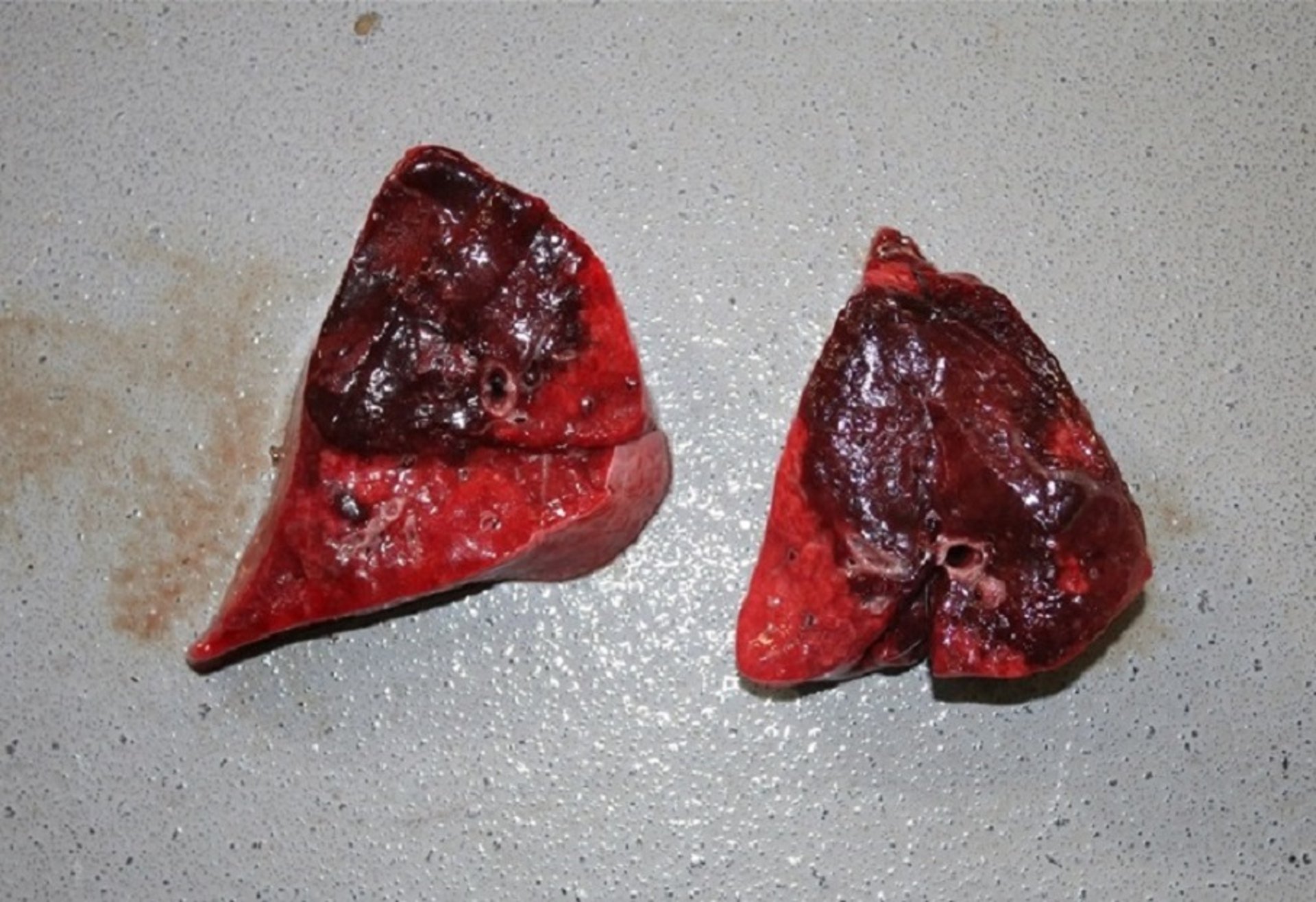
Gross pathology photograph of lung tissue (cut-section) from a goat that died acutely from pneumonia. Culture grew a Mycoplasma sp. Histopathologic findings were suggestive of M haemolytica coinfection, but recent antimicrobial treatment may have prevented its growth on culture.
Courtesy of Dr. Laura Bryan.

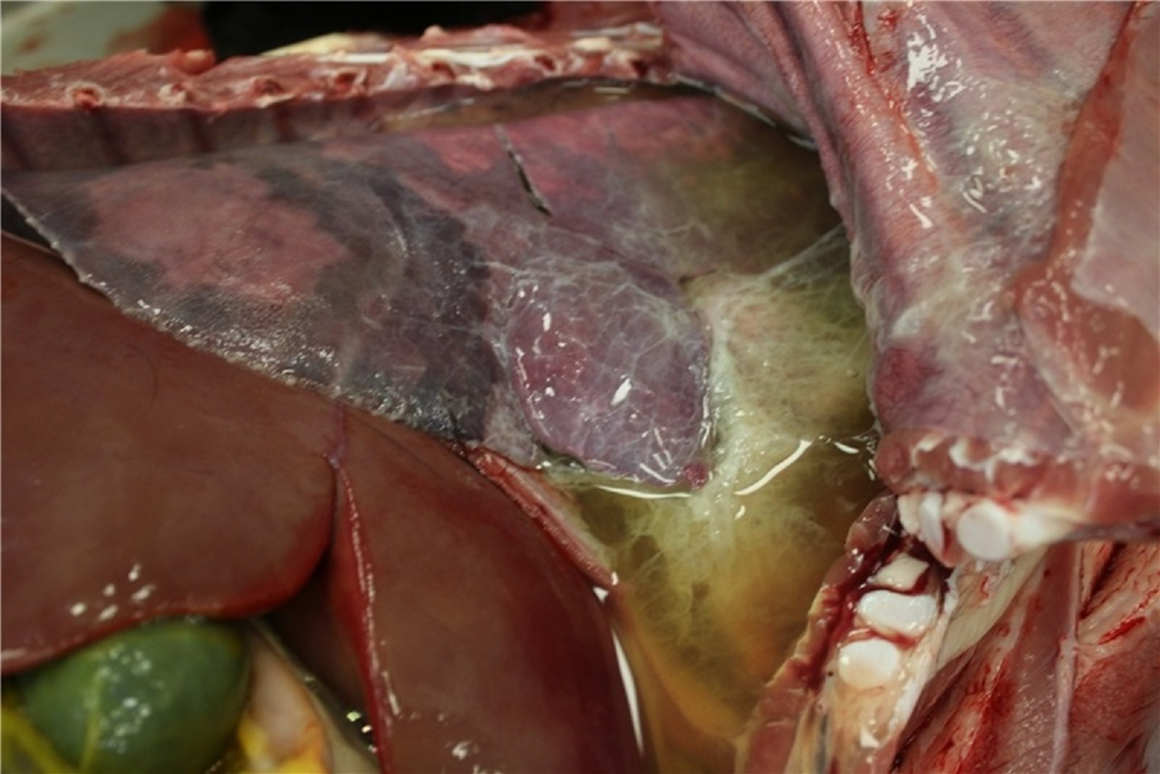
Gross pathology photograph of a goat that died acutely from pneumonia. The thoracic cavity was filled with pleural fluid and fibrin. Pericarditis was also present, and the lung tissue culture grew Mannheimia haemolytica.
Courtesy of Dr. Amanda Anderson.
Subcutaneous ecchymotic hemorrhages occur over the throat and ribs. The lungs are heavy, swollen, and purple-red in peracute cases, and the airways contain blood-stained froth. Cases of longer duration show cranioventral consolidation and fibrinous pleuropneumonia.
Diagnosis of Bacterial Bronchopneumonia in Sheep and Goats
Bacterial culture
At necropsy, testing whether lung tissue sinks (pneumonia) or floats (normal) remains a useful screening test for pneumonias due to Pasteurella or Mannheimia. In acute cases, cultures obtained from tracheal swabs or washes or from lung tissue or associated lymph nodes are diagnostic. Histologic examination is useful, especially if other types of pneumonia (eg, retrovirus interstitial pneumonia in adult sheep and goats) are also suspected. In chronic cases, bacterial cultures may be less rewarding; Pasteurella or Mannheimia may have been the initial problem, but results of cultures taken later may reveal Trueperella pyogenes, a common tertiary pathogen and causative agent of lung abscesses.
Treatment and Control of Bacterial Bronchopneumonia in Sheep and Goats
Antimicrobials
Supportive care
Vaccination
Environmental management
Whenever possible, treatment of Pasteurella and Mannheimia pneumonias should be based on bacterial culture and antimicrobial susceptibility testing, especially in herd or flock outbreaks, when valuable animals are involved, or in acute or chronic cases when initial therapeutic attempts have failed. Susceptibility patterns of isolates may vary considerably. Commonly recommended antimicrobials include oxytetracycline (10 mg/kg, IM or IV, every 24 hours, of non–long-acting product; or 20 mg/kg, IM or IV, once, of a long-acting product); florfenicol (20 mg/kg, IM, every 48 hours; or 40 mg/kg, SC, once); ceftiofur (2.2 mg/kg, IM, every 24 hours for 3–5 days); and tylosin (10–20 mg/kg, IM, every 12–24 hours). Tilmicosin (for use in sheep only), tulathromycin, and other macrolide antimicrobials can also be used; however, they may be considerably more expensive, and tilmicosin poses a high risk of injury and death in cases of human exposure. Acute cases may also benefit from the use of NSAIDs (eg, flunixin meglumine, meloxicam, or ketoprofen) for control of endotoxemia and inflammation. Treatment choices may be influenced by the availability and legality of antimicrobials by region.
Inadequate ventilation, crowding, commingling of animals from various farms (feedlot or sale barn situations), poor nutrition, failure of passive transfer, transportation, and other stresses have all been associated with pneumonia outbreaks. Control and prevention depend on correction of the predisposing factors and prompt response to clinical cases.
For prevention of pasteurellosis, vaccines that incorporate iron-regulated proteins are best. Because these proteins are antigenically similar, they confer cross-protection against multiple serotypes. Breeding ewes require a primary course of two injections 4–6 weeks apart, followed by an annual booster 4–6 weeks before lambing. However, this vaccination regimen provides passive immunity to the lambs for only up to 5 weeks. Lambs can be protected by two doses of vaccine administered from the age of 10 days because colostral antibody does not interfere with the development of active immunity.
Key Points
Young, stressed, and commingled animals are at highest risk for Pasteurella or Mannheimia bronchopneumonia.
Prompt and effective antimicrobial administration is essential for successful treatment.
Prevention relies on good management practices and vaccination.


