

Each veterinary diagnostic laboratory offers a unique set of diagnostic tests that is subject to frequent changes as new and improved tests become available. The protocols for sample collection and submission are therefore also subject to change.
The practitioner and diagnostic laboratory staff must maintain good communication to complete their diagnostic efforts efficiently and provide optimal service to the client. Practitioners must be specific and clear in their test requests. The laboratory staff will provide guidance regarding sample collection and handling protocols, as well as assistance in the interpretation of test results.
Most diagnostic laboratories publish user guidelines with preferred protocols for sample collection and submission; however, the following broad recommendations are fairly standard.
A detailed case history should be included with submitted samples. This information is particularly critical for the interpretation of samples submitted for cytologic and histologic evaluation. The submission form should be protected in a waterproof bag to avoid damage from any fluids in the packaged materials.
A detailed case history should include the following elements:
owner's first and last name
species
breed or size/weight (if breed unknown)
sex
age
animal identification
relevant clinical signs
gross appearance (including size and anatomic location) of lesions
previous treatment, including response to treatment (if any)
results of relevant diagnostic testing, such as CBC, serum biochemical analysis, cytologic and histologic evaluation, and imaging studies
for population health studies, morbidity and mortality in the group
If a zoonotic disease is suspected, this should also be clearly indicated on the submission form to alert laboratory personnel.
Packaging of Samples from Animals for Shipment
The shipment of biological specimens should comply with protocols established by the courier or shipping service used. In some instances, air transport requires compliance with International Aviation and Transportation Association (IATA) regulations for hazardous materials.
These regulations include restrictions on the volume of formalin that can be shipped in a container: no more than 30 mL of free liquid solution in each inner container, and no more than 1 L in the entire outer package. Rather than shipping large volumes of formalin, tissue samples can be fixed in-house for at least 24 hours in an appropriate volume and then transferred into a smaller volume of fresh formalin for shipment.
Shipments with fresh tissue samples are required to be clearly labeled:
UN2814, Infectious substances, affecting humans
UN2900, Infectious substances, affecting animals
UN3373, Biological substances, Category B
Most veterinary diagnostic samples are classified as Category B (an infectious substance not in a form generally capable of causing permanent disability or life-threatening or fatal disease in otherwise healthy humans or animals when exposure to it occurs). If high-risk or reportable diseases are suspected, it is essential to contact state and federal veterinary agencies regarding necessary shipment precautions.
Further details can be found at the International Air Transport Association website and the US Postal Service Postal Explorer website as well as commercial courier websites.
A fundamental approach to packaging samples involves a 3-layer barrier to protect them:
The sample is placed in an appropriate primary container (sealed jar, bag, or tube).
The primary container is then enclosed within a secondary container, which also includes absorbent material.
The secondary container is then placed in the shipping box (tertiary container), which often houses refrigerant or cold packs as well as various cushioning materials (eg, polystyrene foam) to protect the sample. Ideally, the tertiary container is a sturdy polystyrene refrigerator box or a cardboard box lined with a fitted polystyrene lining.
Items such as syringes, obstetrical gloves, and containers without sealable orifices are not suitable for shipment. Under no circumstances should needles, scalpel blades, or any other potentially injurious item (eg, broken microscope slides with sharp edges) be submitted to the lab.
Liquid samples should not ship in plastic bags; instead, a sealable jar should be used. Waterproof markers should be used when labeling specimen bags and containers; at a minimum, patient identification and specimen type (eg, urine, serum, plasma) are critical labeling information.
Coolant materials should be sealed in separate plastic bags to prevent condensation damage. Refrigerant or cold packs should not be placed in direct contact with samples, such as tubes of whole blood, that could suffer adverse effects if frozen in transit.
The suitably protected, completed submission form must be included.
If dry ice is used, this should be noted on the cardboard box label, and the lid should not be sealed with tape. Otherwise, CO2 released from the dry ice could increase pressure and damage the package or contents.
Preparation of Samples from Animals for Histologic Evaluation
Tissues for microscopic examination collected either via biopsy or during necropsy can be critical to obtaining a diagnosis. Immunohistochemical (IHC) and other advanced tests that can be applied to formalin-fixed tissue further reinforce the utility of histologic evaluation as diagnostic technique.
Autolyzed tissues are generally useless for histologic evaluation; prompt necropsy and organ sampling are critical.
Tissue should not be frozen prior to fixation.
Other than CNS tissues, samples collected for histologic evaluation should never be > 1 cm thick (preferably 5–7 mm) and must be immediately placed in ≥ 10 times their volume of phosphate-buffered 10% formalin to ensure adequate fixation.
Tissues should be representative of any lesions present and, in the case of cutaneous punch biopsies and biopsies obtained via endoscopic collection, be centered directly on the grossly visible lesions.
Wedge biopsies or tissue samples collected at necropsy should include some of the apparently normal surrounding tissue; the interface between normal and abnormal tissue may provide key information.
Excisional biopsies of small tumors (< 1.5 cm) may be cut in half. Larger tumors may be sliced like bread so that formalin can penetrate to the face of each slice. Alternatively, several representative samples (7 mm wide, including the interface of normal and abnormal) may be collected.
Rather than shipping large volumes of formalin, tissue samples can be fixed in-house for at least 24 hours in an appropriate volume and then transferred into a smaller volume of fresh formalin for shipment.
Pearls & Pitfalls
|
Submission of tissue that has been stored in formalin is acceptable within any time frame, although quality of sample will degrade over time. Prolonged formalin fixation can adversely affect advanced testing; for example, immunohistochemical testing is not recommended for tissue that has been stored in formalin for > 7 weeks because results may be invalid.
Samples should be shipped in laboratory-approved unbreakable containers with screw-top lids and packed in a manner that prevents spillage or freezing during shipment (see Packaging of Samples for Shipment).
Specific tissues collected at necropsy require additional attention:
Because the GI mucosa decomposes rapidly, short sections of gut collected at necropsy should be opened lengthwise to allow adequate fixation.
If the spinal cord is submitted, the dura mater should be carefully incised lengthwise to permit more rapid penetration to the cord.
Fixing the brain poses a special dilemma, especially if a neuroanatomical location of the lesions within the organ could not be determined antemortem.
Ideally, a whole, intact, fixed brain is required for complete histologic evaluation.
Immersion of the brain for many days in a large volume of formalin is required for adequate fixation; consequently, brains are commonly transported in an only partially fixed state. If the specimen can be shipped by overnight delivery, it may be acceptable to send a chilled, carefully packaged, unfixed brain, which can then be processed at the diagnostic laboratory.
Often, the brain is halved longitudinally and one half is sent unfixed (fresh), properly refrigerated, for microbiological testing, while the other half partially fixes in transit. This method can prove unsatisfactory if a solitary unilateral lesion is involved. Slicing the brain into widths suitable for rapid fixation introduces considerable fixation artifact and should be avoided if possible; fixing the intact (or halved) brain in a large volume of formalin for > 24 hours is preferred.
Preparation of Samples from Animals for Microbiological Testing
Any specific infectious agents that are suspected clinically should be noted on the submission form; some organisms have specific growth requirements (eg, anaerobic culture, special media) that may not be routinely used in laboratories unless the pathogen was cited as a differential diagnosis.
Laboratory techniques and capabilities for microbiological testing vary; however, most tests rely on either the growth or visualization of intact viable organisms or the detection of the nucleic acids and proteins of these pathogens. Therefore, unfixed specimens (tissue, fluid, etc) should be collected aseptically and shipped promptly to avoid degradation.
If microbial culture of a fluid specimen such as urine or a cavitary effusion is a possibility, a separate aliquot of the fluid in a sterile, additive-free container should be submitted. Tubes containing EDTA or other preservatives are generally ideal for cytologic evaluation but unacceptable for culturing.
If PCR assays are to be performed, it is particularly important to avoid cross contamination between multiple animals in a submission; this principle applies to tissues, fluids, and even dissection instruments.
Furthermore, swabs for PCR analysis should not be placed in agar or charcoal-based transport media, and calcium alginate swabs should be avoided. Instead, sterile cotton- or polyester-tipped applicators should be shipped in a tube, either dry (without transport media) or with a few drops of sterile saline solution or viral transport media, depending on the individual laboratory's requirements.
Requirements for PCR assay can vary widely by specific PCR test, and consulting the recipient laboratory before obtaining the sample is strongly encouraged.
Some test protocols may permit pooling of organ specimens from an individual; however, it is generally preferable that each tissue be collected into separate, clearly labeled sterile bags or tubes for shipping. Gut samples must never be pooled in a container with other tissue samples.
Tissues and fluids for most microbiological assays may be frozen before shipment, but freezing is generally undesirable if samples can be chilled and delivered directly to the laboratory within 24 hours after collection.
Exceptions to this rule include analysis for certain toxins, such as those of Clostridium perfringens and C botulinum, in which degradation of the toxin must be prevented by prompt freezing after collection. Adequate coolant should be provided so that samples remain chilled (or frozen) until they reach the laboratory.
Fecal samples for parasitological testing should be submitted chilled in appropriately sealed containers. Freezing may have little impact on routine flotation or sedimentation tests but will negate the possibility of Baermann analysis for nematode larvae. Ectoparasites or nematodes being submitted for identification should be submitted in vials containing 70% alcohol.
Preparation of Samples from Animals for Toxicological Testing
If a known toxin is suspected, a specific analysis should always be requested—laboratories cannot just “check for poisoning.” A complete description of clinical and epidemiological findings may help differentiate poisoning from infectious diseases that mimic poisoning.
Pearls & Pitfalls
|
The most critical samples to be collected are generally the following:
gastric contents
liver
kidney
whole blood
plasma or serum
urine
There are also some exceptions, such as the collection of cerebral tissue for cholinesterase analysis.
For some investigations, the diagnosis requires analysis of feed or water. If there is doubt regarding sample submission procedures, the laboratory should be contacted.
Freezing is critical to prevent the degradation of a few analytes, such as cholinesterase, zinc phosphide, and sodium fluoroacetate (compound 1080). However, for most analytes, overnight shipping of chilled specimens will suffice.
The containers for packing and transporting specimens should be free of chemicals. Plastic containers (eg, jars) are ideal, and jars with metal tops should be avoided. Samples should be packed individually and all containers labeled.
If legal action is a possibility, all containers for shipment should be either sealed so that tampering can be detected or hand-carried to the laboratory and a receipt obtained. The chain of custody must be accurately documented.
If feed or water is suspected as the source of poisoning, chilled samples of these along with any descriptive feed tag should accompany the tissue samples. If at all possible, a representative composite sample of the feed should be submitted from the suspect lot or shipment (ie, aliquots from the top, middle, and bottom of the feed container).
Preparation of Samples from Animals for Hematological Testing
Routine hematological tests require anticoagulated whole blood filled to the appropriate fill volume for the specimen collection tube. Overfilling or underfilling of tubes can cause unreliable analytic results due to an incorrect blood-additive ratio.
Ideally, several blood smears should be included. Blood smears should be prepared immediately after the sample has been collected in order to minimize artifacts, including altered erythrocyte morphology, platelet clumping, and general cell deterioration.
Anticoagulated blood should be kept refrigerated; blood smears should be stored at room temperature. EDTA is the anticoagulant of choice for mammalian CBCs.
EDTA is acceptable for most, but not all, nonmammalian species. Blood from stingrays, some bony fish, and some reptilian and avian species undergoes hemolysis when collected into EDTA, so heparin is commonly used for these species.
Blood for coagulation testing should be collected into a citrate (blue-top) tube containing trisodium citrate (3.2% or 3.8%, as specified by the reference laboratory). It is imperative that the tube be filled to the appropriate fill volume because underfilling can cause falsely prolonged clotting times.
For plasma-based assays, citrated plasma typically must be separated from whole blood within 6 hours after collection for measurement of prothrombin time (PT) and activated partial thromboplastin time (aPTT). Plasma is separated by centrifugation at 1,500g for 15 minutes, after which the plasma is transferred to an additive-free plastic secondary tube using a disposable plastic pipette.
Plasma should ideally be analyzed within 1 hour after separation. However, many samples for coagulation testing are sent to reference laboratories for analysis, and adherence to laboratory-specific guidelines pertaining to specimen storage is advised.
Freezing of whole blood causes cell lysis and gross hemolysis, which interfere with testing; freezing of whole blood (during storage or specimen transport) must be avoided.
Preparation of Samples from Animals for Serum Biochemical Analysis
Anticoagulants present in plasma may result in spuriously increased or decreased test analyte values (eg, samples contaminated with potassium-EDTA have artifactually elevated potassium concentrations and decreased calcium concentrations); therefore, by default, serum should be submitted.
However, in some instances, heparinized plasma may be acceptable or, possibly, required. Consultation with the lab before collecting the specimen is recommended for any nonroutine tests or species.
Because lipemia can interfere with a number of chemistry tests for dogs and cats, food withholding is advisable for 12 hours before samples are collected unless there is a potential medical contraindication, such as with pediatric patients or toy dog breeds.
For serum samples, the blood should be drawn into a serum (red-top) tube or serum separator (tiger-top) tube. The sample should be held at room temperature for 20–30 minutes to allow complete clot formation and retraction. Incomplete clot formation may cause the serum to gel due to latent fibrin formation.
The clot should be separated from the glass by gently rimming the tube walls with an applicator stick to loosen the clot. The sample should then be centrifuged at moderate speed (~450 rpm) for 10 minutes.
If the sample has been collected into a serum separator tube, centrifugation will cause a layer of polymer gel to lodge between the packed cells and the serum. The gel layer should be inspected to ensure the integrity of the barrier, and repeated centrifugation is recommended if there is a visible crack in this layer.
If a serum (red-top) tube has been used, the serum should be removed and transferred to a clean tube to minimize artifacts (eg, glycolysis in the cellular component of the blood sample may result in an artifactually decreased glucose concentration). Serum should be refrigerated or frozen until analyzed. Delays in analysis may adversely affect the results for a number of analytes.
Rough handling of the sample or incomplete separation of erythrocytes from serum may promote hemolysis, which can interfere with certain tests. For chemistry panels, 0.5 mL of serum is typically preferred, although minimum laboratory requirements may vary and should be consulted for serum volumes < 0.5 mL.
Preparation of Samples from Animals for Urinalysis
Urine samples should be clearly labeled with specimen type because urine and serum may be grossly indistinguishable.
Noting the method through which the urine was obtained (free catch, catheterization, cystocentesis, etc) can be helpful for interpretation of findings; for example, free catch samples may have bacterial contaminants from the external genitalia and perineum, and catheterized samples often have greater numbers of transitional epithelial or urothelial cells due to mechanical exfoliation from passing of the catheter.
Clear specimen containers can facilitate gross examination of the urine. However, if urine is not evaluated within 30 minutes after collection, care must be taken to protect it from exposure to ultraviolet light, which causes degradation of urine constituents, including bilirubin.
Lids must be securely screwed on to prevent leakage, evaporation, or volatilization of ketones (if present). Containers must not be reused because traces of detergent can interfere with chemical analysis and bacterial contaminants can proliferate during specimen storage and transit.
Urine should be stored and shipped chilled, although it should be noted that crystal precipitation can occur. Urine is a caustic environment that causes rapid degradation of formed elements, including cells.
If urinary tract disease reflected by the sediment examination (eg, inflammation, infection, proliferative process involving neoplasia) is suspected, cytologic smears prepared from fresh urine sediment should be submitted along with the urine sample. Cytologic smears are air-dried and are prepared as described in the Samples for Cytologic Evaluation section.
As with other fluid samples, volume requirements apply to samples for urinalysis. The preferred volume is generally 5–6 mL, although lesser volumes may be accepted, depending on the lab.
Preparation of Samples from Animals for Serologic Testing
Serologic testing generally requires serum, but plasma is often satisfactory. Samples should be collected as described for serum biochemical analysis and should always be free of hemolysis.
In some instances, paired samples may be required for an adequate diagnosis. The acute sample should be collected early in the course of the disease and frozen. The convalescent sample should be collected 10–14 days later, and both samples should be forwarded to the laboratory at the same time.
Preparation of Samples from Animals for Cytologic Evaluation
For superficial mass effects, sampling by fine needle (ie, aspiration or coring/"woodpecker" technique) is typically of higher diagnostic yield and more representative of the primary lesion than samples obtained via surface impression. Impression smears are prone to contamination with debris and flora of the skin or environment, or may represent only secondary inflammation or infection.
Aspirated material must be spread into a monolayer (ie, approximately half the cells touch but do not overlap) after application to the slide (see and slides).

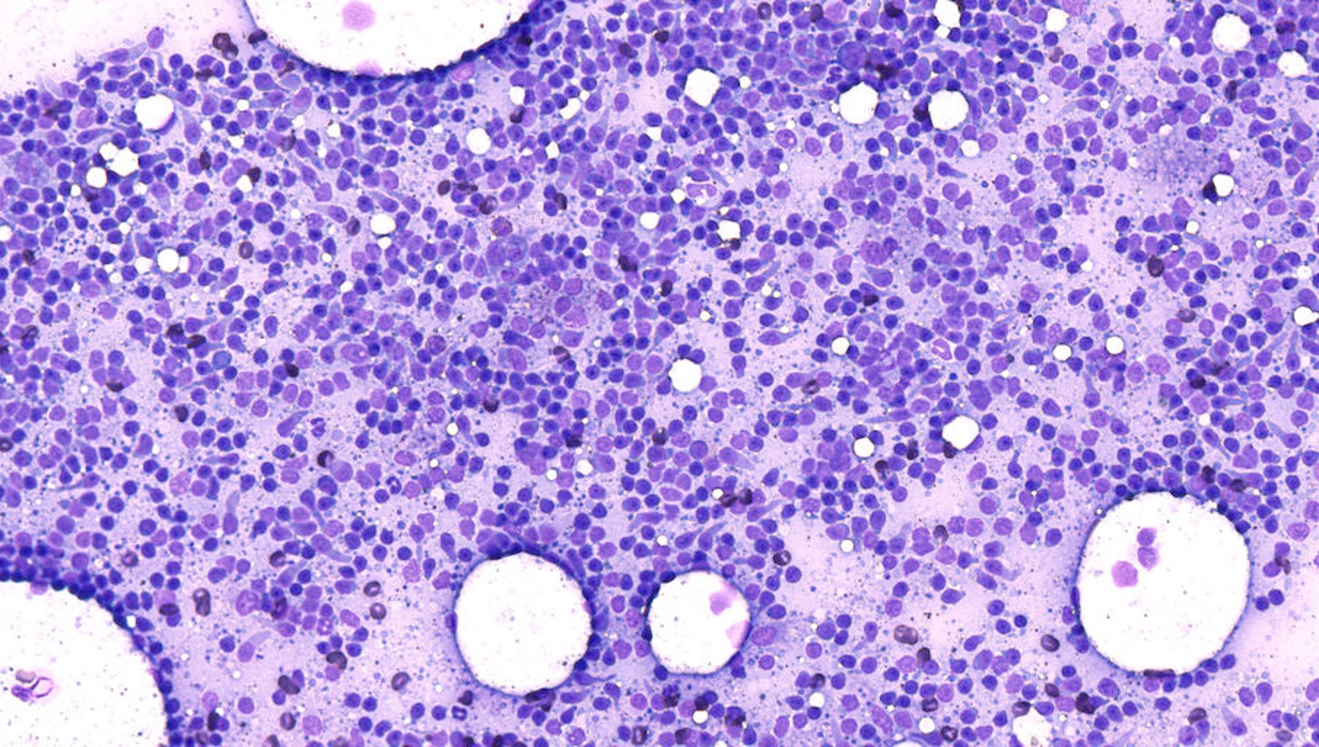
Photomicrograph of an area of the monolayer of a cytologic smear of a lymph node aspirate. The lymphocytes are sufficiently spread to accurately appreciate their size and other morphological characteristics. Approximately half the cells are touching, but no appreciable overlapping is noted. The white circles in the background represent nonstaining lipid material from perinodal adipose tissue. Aqueous Romanowsky stain; original magnification, 200X.
Courtesy of Dr. Rebekah Gunn-Christie.

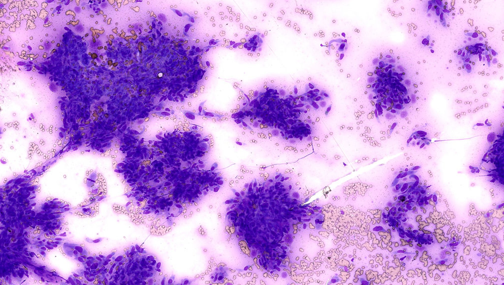
Photomicrograph of an area of the monolayer of a cytologic smear showing proliferation of mesenchymal cells. Although dense groups of cells are unavoidable, there are many mesenchymal cells that are sufficiently spread for evaluation. The monolayer characteristic of the smear is readily apparent by looking at the erythrocytes in the background, where approximately half are touching but no appreciable overlapping is present. Aqueous Romanowsky stain; original magnification, 200X.
Courtesy of Dr. Rebekah Gunn-Christie.
The preferred spreading method involves gentle horizontal smearing using a second slide that is placed on top of the material after it is deposited on the bottom slide (see ). This technique is superior to the vertical pull-apart technique (see ).

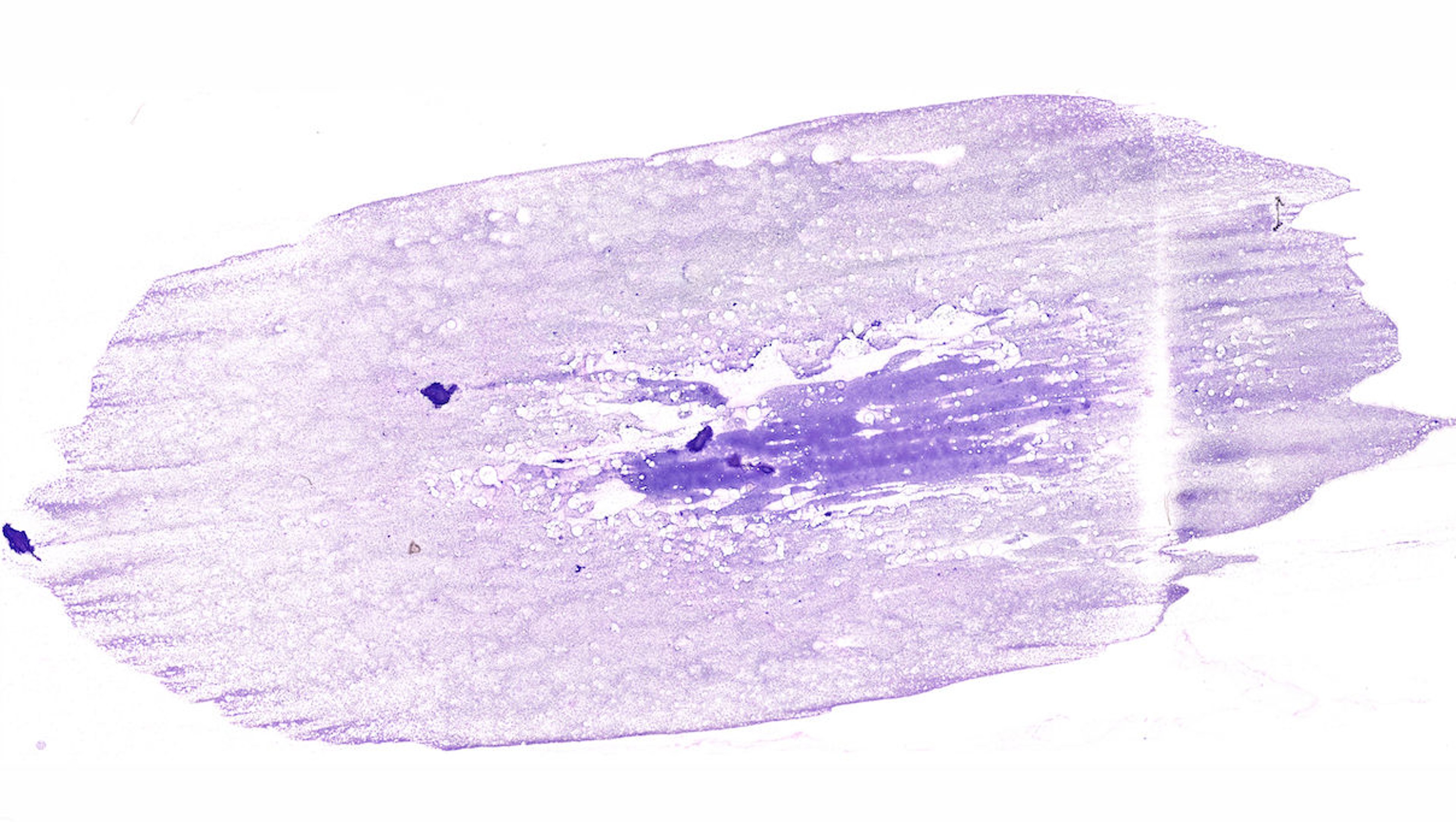
Subgross photograph of a cytologic smear prepared with gentle horizontal smearing using a second slide that was placed on top of the material after it was deposited on the bottom slide. Much of the diagnostic material is present in a monolayer and free from excessive cytolysis. Aqueous Romanowsky stain; original magnification, 2X.
Courtesy of Dr. Rebekah Gunn-Christie.

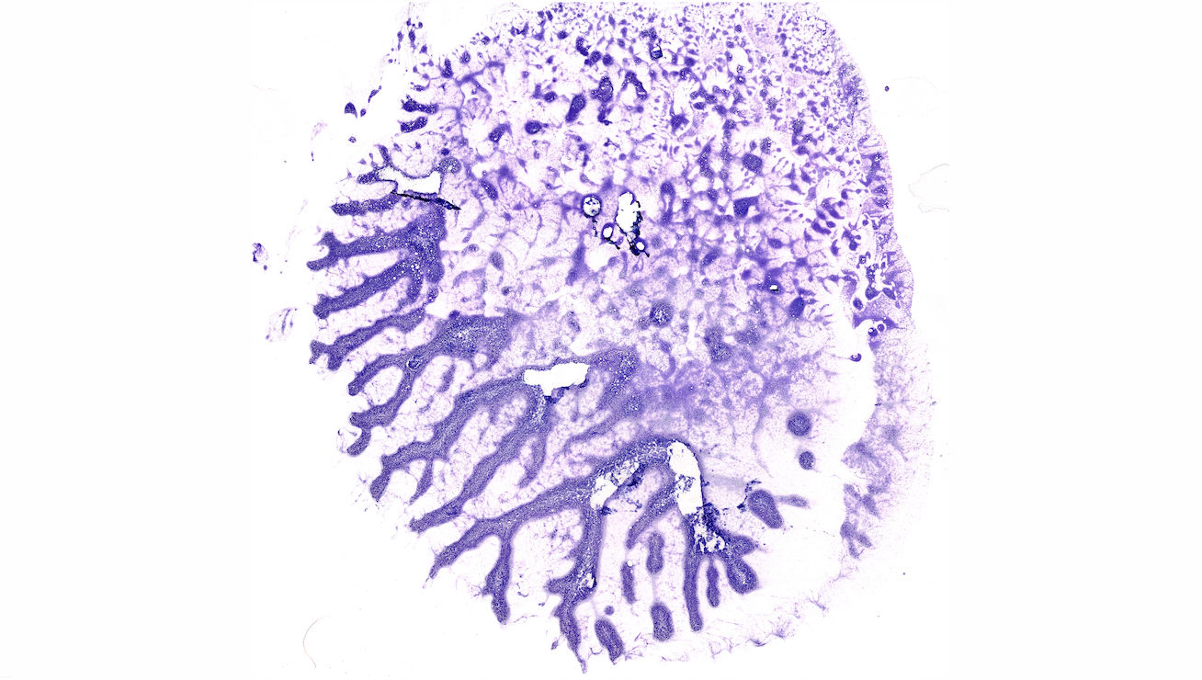
Subgross photograph of a cytologic smear prepared with vertical pull-apart technique. Most areas of cellular material are characterized by excessive cytolysis or specimen density, with very few areas amenable to cytologic evaluation. Aqueous Romanowsky stain; original magnification, 2X.
Courtesy of Dr. Rebekah Gunn-Christie.
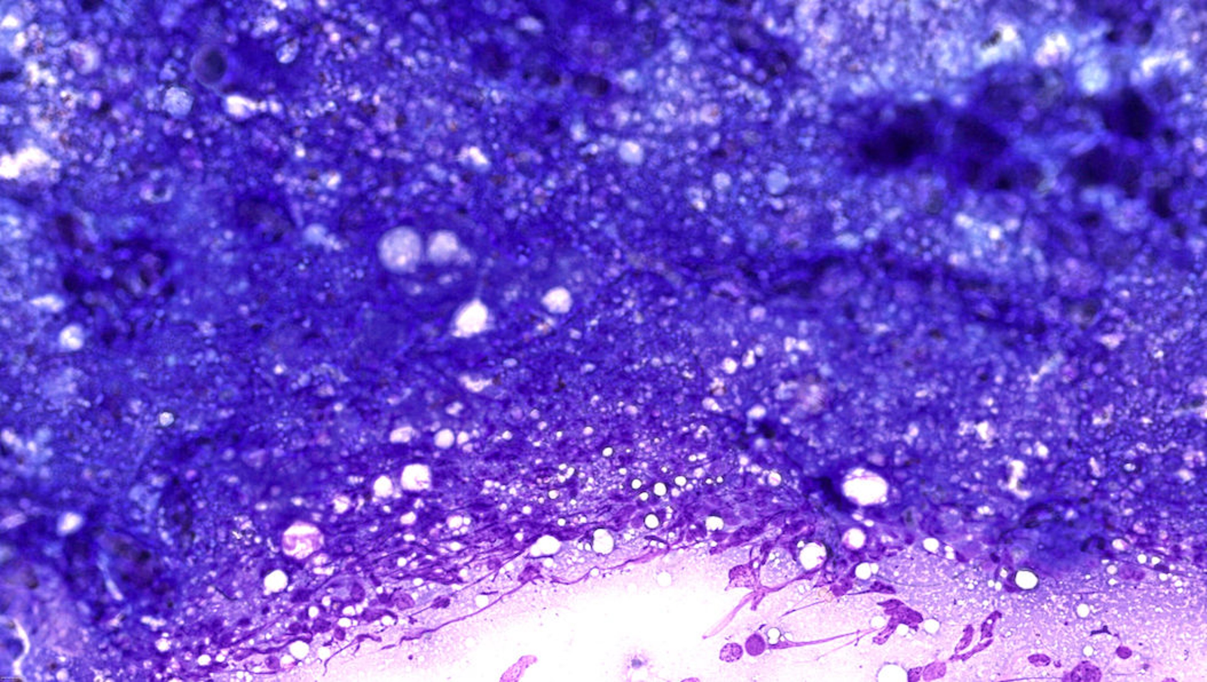
Excessive specimen density and cytolysis inhibit the ability to adequately evaluate cells in cytologic specimens (see and slides; see also slide). Only intact cells can be evaluated cytologically because cytolysis introduces artifacts that can mimic malignancy, such as enlargement of nuclei and increased prominence of nucleoli.


Subgross photograph of a cytologic smear showing excessive cellular density. The “fractures” in the cellular material are a result of excessive specimen density. Aqueous Romanowsky stain; original magnification, 2X.
Courtesy of Dr. Rebekah Gunn-Christie.

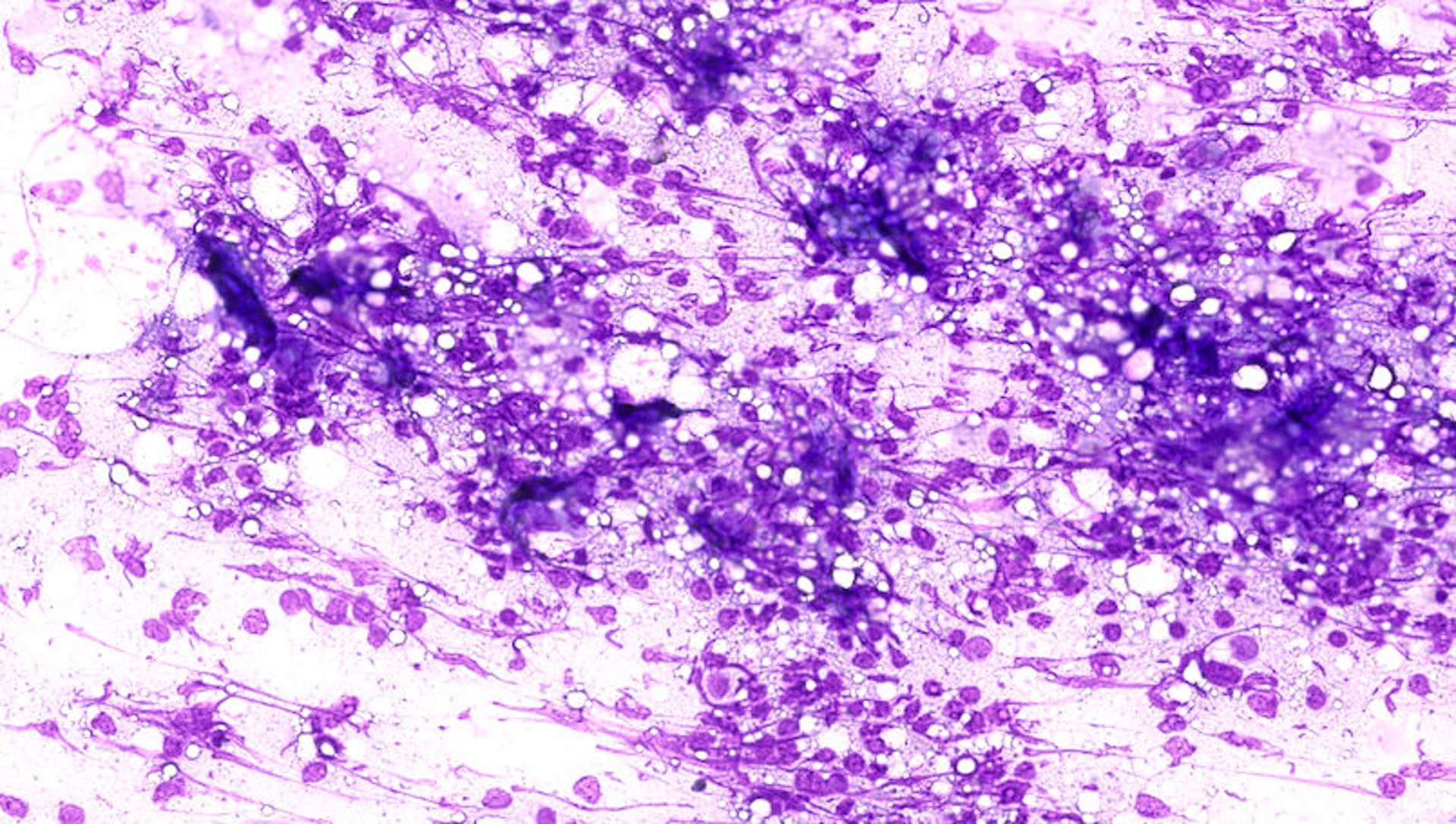
Photomicrograph of a cytologic smear showing excessive cellular density. The cells are too tightly packed to evaluate individual cells. Aqueous Romanowsky stain; original magnification, 400X.
Courtesy of Dr. Rebekah Gunn-Christie.

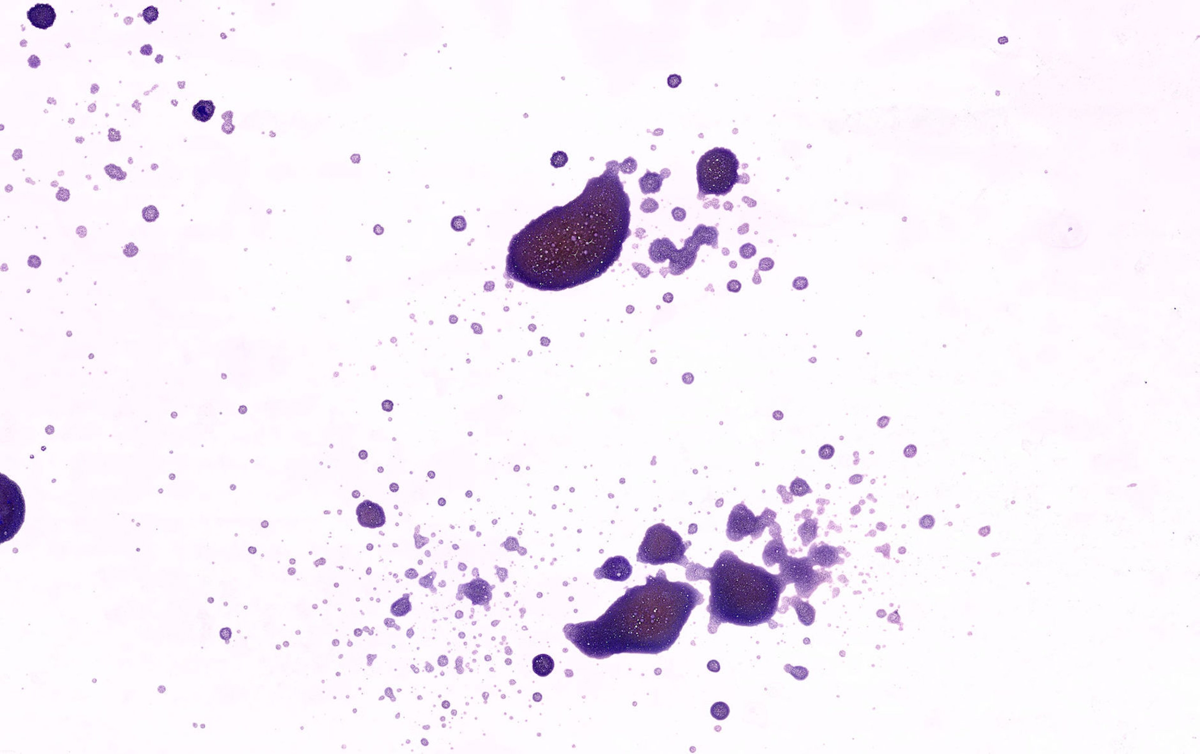
Photomicrograph of a cytologic smear showing excessive cytolysis. Nearly all cells in this field are lysed, as represented by bare and streaming nuclei devoid of surrounding cytoplasm. Aqueous Romanowsky stain; original magnification, 400X.
Courtesy of Dr. Rebekah Gunn-Christie.
Both the pull-apart technique and the lack of any effort to spread the material (ie, simple air drying of droplet-form deposited biological material) are prone to excessive specimen density and cytolysis, and may be nondiagnostic (see slide).

Subgross photograph of a cytologic smear where the specimen was not spread after it was applied to the microscope slide. The preparation lacks a monolayer and is affected by excessive specimen density throughout areas of cellular material. Aqueous Romanowsky stain; original magnification, 2X.
Courtesy of Dr. Rebekah Gunn-Christie.
Samples with a fluidic component can be spread in the same manner as blood smears. Highly cellular fluids may be smeared directly; fluids of low cellularity should be centrifuged to concentrate the cells.
Slides should be stored at room temperature and protected from dust as well as excessive humidity and prolonged exposure to light. It is important to physically label slides with patient identification as well as the source of the cytologic sample; labeling only slide boxes or cartons is unreliable because slides may be separated from the containers or containers reused.
Samples containing excessive ultrasound or lubricant gel may be nondiagnostic because this material is microscopically opaque and obscures cells as well as causing poor cellular stain uptake.
Other practical considerations for preparation of cytologic samples include always using fresh slides, avoidance of coverslips and acetate tape, and ensuring that cellular material is deposited towards the center of a single side of the slide (see ).
As digital scanning of slides becomes more prevalent in diagnostic labs, it is not possible for a pathologist to simply flip over the slide on their microscope to focus on the material on the opposite slide. In addition, if the material on the two sides overlaps, it is possible that the net effect will be material that is too thick to adequately assess.


Subgross photograph of a cytologic smear with cellular material on both sides of the slide. Aqueous Romanowsky stain; original magnification, 2X.
Courtesy of Dr. Rebekah Gunn-Christie.
Exposure of blood and cytologic smears to formalin or formalin fumes (including proximity to sealed jars of formalin) will interfere with adequate staining and evaluation, potentially resulting in a nondiagnostic specimen. These slide preparations must never be submitted to the laboratory in the same package as formalin-fixed tissues.
Air-dried, unfixed smears are usually appropriate for immunocytochemical (ICC) staining; however, in some instances, shipping samples in tubes containing transport media is recommended. If special stains including ICC are anticipated, the laboratory should be contacted prior to submission to ensure their laboratory-specific protocol is followed.
Preparation of Samples from Animals for Fluid Analysis
Analysis of cavitary (peritoneal, pleural, and pericardial) effusions and other fluids (eg, synovial fluid, CSF) includes determination of protein content, total erythrocyte and WBC counts, and cytologic evaluation. Other tests may be performed depending on the source or appearance (eg, chylous fluid) of the effusion.
A sample of fluid should be collected into an EDTA (purple-top) tube for routine analysis. A second sample should be collected into a serum (red-top) tube if microbial culture and antimicrobial susceptibility testing or biochemical analyses are anticipated. These samples should be shipped chilled but not frozen.
Cytologic evaluation without measurement of other parameters (eg, total protein concentration, cell counts) is usually acceptable for mass effects characterized by a fluidic component.
Smears for cytologic evaluation should be prepared from a drop of the fluid immediately after the sample has been collected to minimize cell deterioration and other in vitro artifacts. Cytologic slides should be labeled with patient ID, source, and type of preparation (eg, direct, concentrated/sediment) and fluid and then submitted to the lab.
Samples of CSF should be collected into small EDTA tubes and shipped immediately with high priority; the cytologic value of CSF samples degrades rapidly, and the low cellularity makes examination of direct smears unrewarding. If sufficient CSF is available, then a serum (red-top) tube sample may be useful for serologic evaluation or culture attempts.
Preparation of Samples from Animals for Flow Cytometry
Flow cytometry can be performed on samples with an inherently fluidic composition (whole blood, cavitary effusions) and on organ (eg, lymph node, bone marrow) aspirates collected into fluid medium. Intact, well-preserved cells are necessary for accurate results. Consequently, samples 48 hours old or less are usually needed to ensure cell viability.
Specimen volumes can vary, depending on type of sample (eg, whole blood versus organ aspirate), and the lab should be consulted before collection of the specimen.
The lab may have additional recommendations pertaining to sample collection and submission. For example, depending on specimen transit times, it may be advisable to submit samples early in the week to avoid delays spanning the weekend.
Preparation of Samples for Genetic Analysis in Animals
Tests based on the detection of specific genetic features range from karyotype analysis to the identification of specific genes.
The laboratory offering the test should be contacted to determine the specifics of sample collection and handling; required samples range from hair to skin or blood. Many blood-based analyses require collection into acid-citrate-dextrose (yellow-top) tubes and overnight shipment of chilled tubes to the laboratory.
Tissue samples for genetic analysis should be unfixed and shipped immediately after collection. As with most molecular techniques, aseptic collection and the prevention of cross contamination between samples is critical for reliable test results.
Key Points
Communication between the clinician and the veterinary diagnostic laboratory is critical to assure that the appropriate test and sample are used and that results are interpreted correctly. Communication is particularly crucial when using a new laboratory or ordering nonroutine testing.
Samples should be clearly labeled with waterproof marker on appropriate primary containers, then shipped to the laboratory using the "three-layer barrier" format, including a suitably protected submission form detailing case features and specific test requests.
For More Information


