

Duck viral enteritis is an acute and often severe disease of both wild and domestic waterfowl that is induced by duck viral enteritis virus (Mardivirus anatidalpha1). Clinical signs include droopiness and watery or bloody diarrhea. Lesions include generalized hemorrhages and necrosis of the GI mucosa and liver. Prevention includes biosecurity and avoiding contact between domestic or captive waterfowl and free-living waterfowl. Some producers in the US, Canada, and Europe use live, attenuated virus vaccines to immunize ducklings and breeder ducks.
Duck viral enteritis (DVE) is an acute, highly contagious disease of ducks, geese, and swans of all ages, characterized by sudden death, a high mortality rate (particularly among older ducks), and hemorrhages and necrosis in internal organs.
Etiology and Pathogenesis of Duck Viral Enteritis
Duck viral enteritis virus (DVEV), the causative agent of DVE, is a member of the family Orthoherpesviridae, subfamily Alphaherpesvirinae, and genus Mardivirus (the virus species Mardivirus anatidalpha1). Field strains of this virus display differences in virulence; however, all strains seem to be immunologically identical.
DVEV induces vascular damage, especially in smaller blood vessels (venules and capillaries). This damage leads to generalized hemorrhages and progressive degenerative changes in parenchymatous organs.
Apoptosis and necrosis of lymphocytes induced by DVEV may result in lymphoid depletion and possibly immunosuppression.
An immunosuppressive state induced by DVE may explain the presence of secondary bacterial infections due to Pasteurella multocida, Riemerella anatipestifer, and Escherichia coli, which frequently occur in natural outbreaks of DVE in ducklings.
Like other herpesviruses, DVEV may undergo latency, and the trigeminal ganglion seems to be a latency site for the virus. Recovered birds may carry the virus in its latent form, and viral reactivation may be the cause of outbreaks in susceptible wild and domestic ducks.
Epidemiology of Duck Viral Enteritis
The virus that causes DVE is transmitted primarily by direct contact between infected and susceptible ducks or by indirect contact of susceptible ducks with a contaminated environment. Water seems to be a natural route of transmission. Outbreaks of DVE are frequent in captive or domestic duck flocks with access to bodies of water that are also occupied by free-living waterfowl.
Parenteral, intranasal, or oral administration of infected tissues can establish experimental DVEV infection.
A carrier condition is suspected in wild birds. Recovered birds become latently infected carriers and may shed the virus periodically. Captive-reared and released mallards (Anas platyrhynchos) latently infected with DVEV may transmit the virus to native waterfowl when they are released.
DVE has been reported in domestic and wild waterfowl in Europe, Asia, North America, and Africa. Outbreaks of DVE have resulted in limited to serious economic losses on domestic duck farms and sporadic die-offs (ranging from limited to massive) in wild waterfowl. In the US, considerable economic losses due to DVE have been reported in the concentrated duck-producing areas of Long Island, New York.
Members of the family Anatidae (ducks, geese, and swans) are the natural hosts for the virus that causes DVE. Anatids vary in their susceptibility to the virus; Muscovy ducks are the most susceptible. However, naturally occurring infections have been reported in a variety of domestic ducks, including Pekins, Khaki Campbells, Indian Runners, and some mixed breeds. The infection has not been reported in other avian species, mammals, or humans, and it does not pose a zoonotic risk.
The age at infection by DVEV ranges from 7 days to adulthood.
The incubation period for DVE is 3–7 days.
The mortality rate of DVE varies from 5% to 100%, depending on the virulence of the infecting viral strain.
Clinical Findings of Duck Viral Enteritis
The first clinical sign of DVE is often sudden death, commonly associated with a persistently high mortality rate.
Adult ducks with DVE usually die in higher proportions than young ones, increasing the economic importance of the disease. Sick birds are unable to stand, and they show signs of weakness and listlessness (see ).

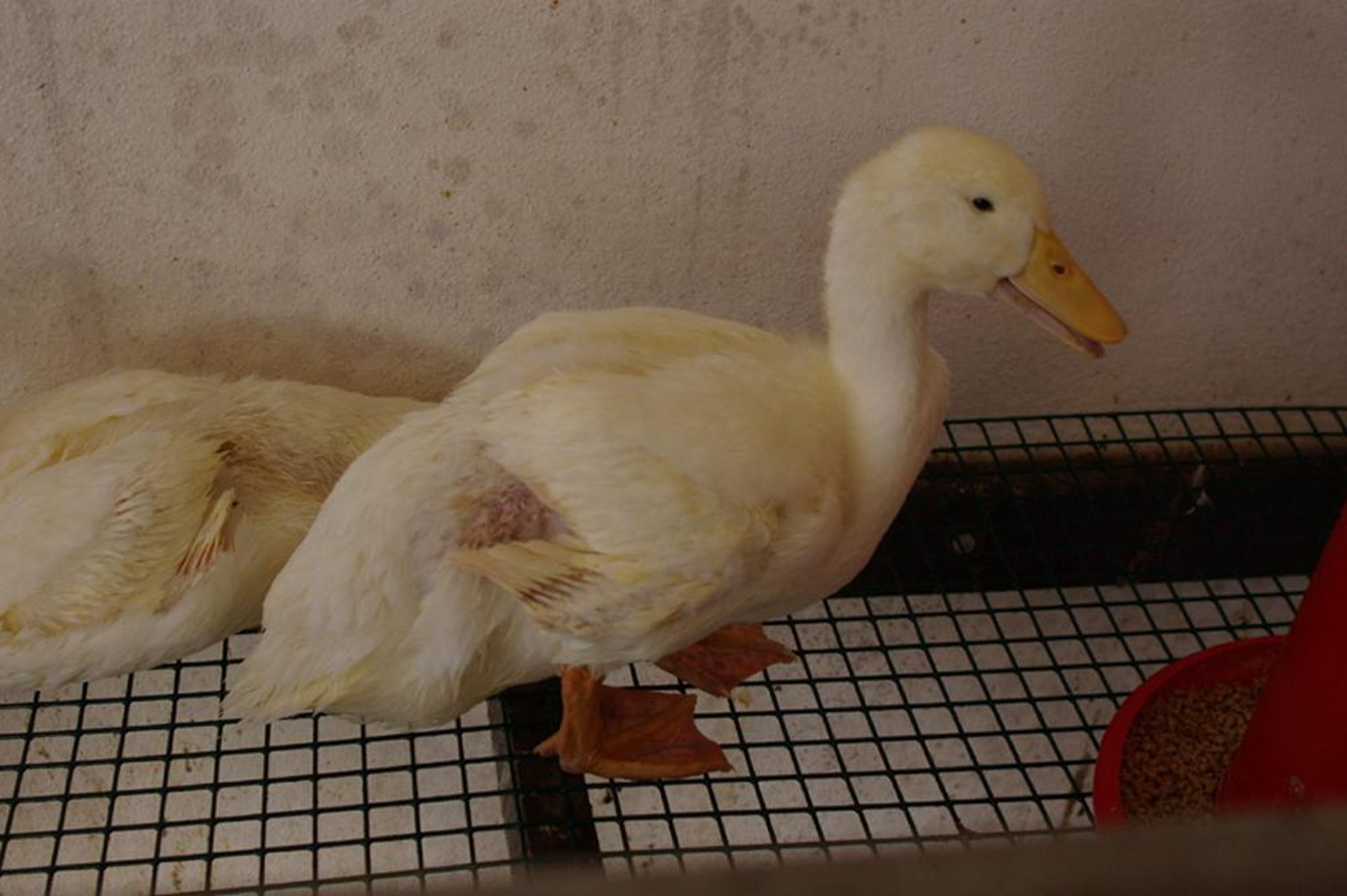
Courtesy of Dr. Alejandro Banda.
Photophobia, inappetence, extreme thirst, droopiness, ataxia, nasal discharge, soiled vents, and watery or bloody diarrhea may be observed in ducks with DVE.
Adult ducks with DVE may die in good body condition. In contrast, ducklings frequently become dehydrated and lose weight, and they may have blue beaks and blood-stained vents. Dead males may have prolapsed penises. In laying flocks, egg production may drop sharply.
Lesions
Lesions due to DVE are indicative of DIC and necrosis of the mucosa and submucosa of the GI tract and lymphoid tissues. Damage to blood vessels throughout the body induces hemorrhages in various tissues or the presence of free blood in body cavities. Petechial and ecchymotic hemorrhages on the heart (“paint brush” appearance), liver, pancreas, mesentery, and other organs are characteristic.
Mucosal eruptions, found in DVE cases in the oral cavity, esophagus, ceca, rectum, and cloaca, undergo progressive alteration during the course of the disease. Macular hemorrhages initially develop into elevated, yellowish, crusted plaques and organize into green, superficial scabs, which may coalesce into large, patchy, diphtheritic membranes. The mucosal lesions align parallel with the longitudinal folds in the esophagus (see ). Diphtheritic esophagitis may commonly occur in swans.
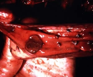
Lumina of the intestines and gizzard are often filled with blood in birds with DVE (see and images). The liver is enlarged, pale copper in color, and may have pinpoint surface hemorrhages mixed with white necrotic foci (see ). The pancreas may have petechiation and multifocal necrosis.
The lymphoid organs in birds with DVE are severely affected. The spleen, which is an active site for viral replication, may be enlarged and darkened because of congestion (see ). The thymic lobes may have petechiation (see ), and thymic atrophy has been reported with some strains. The cloacal bursa (bursa of Fabricius) may be severely congested or hemorrhagic.

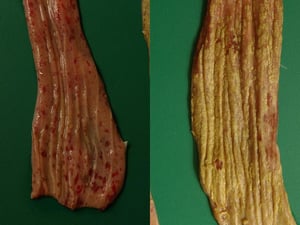
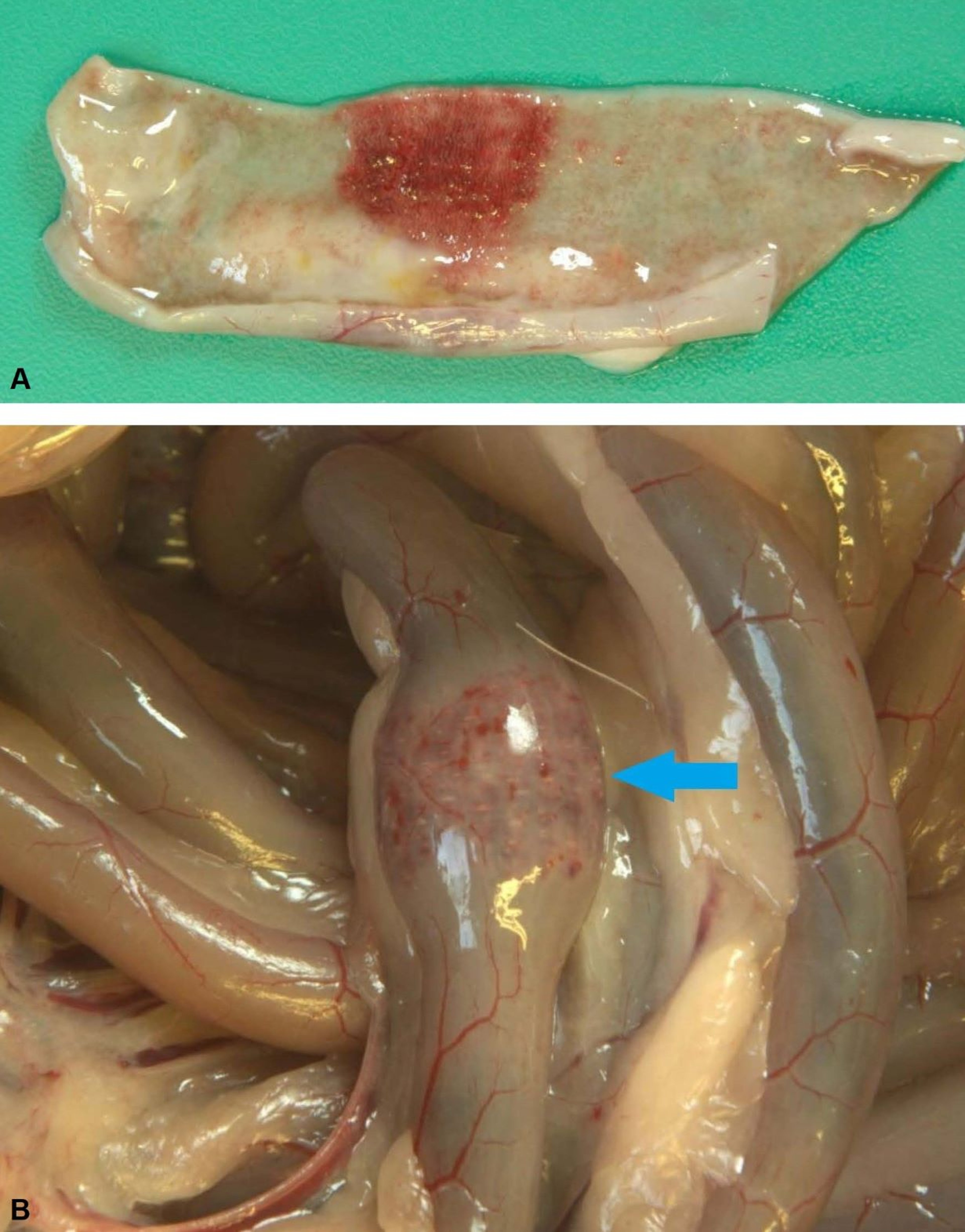
Esophageal tissue taken from a duck with duck viral enteritis. The mucosa of the esophagus shows ecchymotic hemorrhages and ulcers in acute cases (left). In more chronic cases (right), the lumen becomes lined with a yellowish-white membrane or, in some cases, the entire mucosa may be sloughed.
Esophageal tissue taken from a duck with duck viral enteritis. The mucosa of the esophagus shows ecchymotic hemorrhages
Courtesy of Dr. Alejandro Banda.

Multifocal ulcerations on the mucosal surface of the intestine in duck viral enteritis.
Multifocal ulcerations on the mucosal surface of the intestine in duck viral enteritis.
Courtesy of Dr. Jean Sander.

This proventriculus and gizzard from a duck with duck viral enteritis show hemorrhagic contents. The proventriculus mucosa is swollen because of inflammation.
This proventriculus and gizzard from a duck with duck viral enteritis show hemorrhagic contents. The proventriculus muc
Courtesy of Dr. Alejandro Banda.

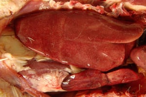
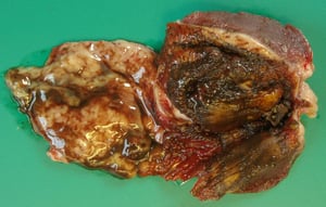
This duck liver is enlarged as a result of hepatitis. The liver color is pale yellowish to tan, with multiple pale foci that correspond to areas of necrosis. In late stages of the disease, a pale copper color and diffuse petechiation of the liver's surface are clinical signs of duck viral enteritis, and pale areas are indicative of focal necrosis.
This duck liver is enlarged as a result of hepatitis. The liver color is pale yellowish to tan, with multiple pale foci
Courtesy of Dr. Alejandro Banda.

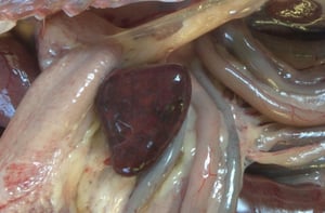
This photograph of the internal organs of a duck with duck viral enteritis shows an enlarged spleen with areas of congestion and focal areas of lymphoid depletion and necrosis (pale areas).
This photograph of the internal organs of a duck with duck viral enteritis shows an enlarged spleen with areas of conge
Courtesy of Dr. Alejandro Banda.

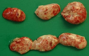
This thymus, taken from a duck with duck viral enteritis, shows mottling with multiple petechiae and necrotic focal areas.
This thymus, taken from a duck with duck viral enteritis, shows mottling with multiple petechiae and necrotic focal are
Courtesy of Dr. Alejandro Banda.
Esophageal tissue taken from a duck with duck viral enteritis. The mucosa of the esophagus shows ecchymotic hemorrhages and ulcers in acute cases (left). In more chronic cases (right), the lumen becomes lined with a yellowish-white membrane or, in some cases, the entire mucosa may be sloughed.
Esophageal tissue taken from a duck with duck viral enteritis. The mucosa of the esophagus shows ecchymotic hemorrhages
Courtesy of Dr. Alejandro Banda.
Multifocal ulcerations on the mucosal surface of the intestine in duck viral enteritis.
Multifocal ulcerations on the mucosal surface of the intestine in duck viral enteritis.
Courtesy of Dr. Jean Sander.
This proventriculus and gizzard from a duck with duck viral enteritis show hemorrhagic contents. The proventriculus mucosa is swollen because of inflammation.
This proventriculus and gizzard from a duck with duck viral enteritis show hemorrhagic contents. The proventriculus muc
Courtesy of Dr. Alejandro Banda.
This duck liver is enlarged as a result of hepatitis. The liver color is pale yellowish to tan, with multiple pale foci that correspond to areas of necrosis. In late stages of the disease, a pale copper color and diffuse petechiation of the liver's surface are clinical signs of duck viral enteritis, and pale areas are indicative of focal necrosis.
This duck liver is enlarged as a result of hepatitis. The liver color is pale yellowish to tan, with multiple pale foci
Courtesy of Dr. Alejandro Banda.
This photograph of the internal organs of a duck with duck viral enteritis shows an enlarged spleen with areas of congestion and focal areas of lymphoid depletion and necrosis (pale areas).
This photograph of the internal organs of a duck with duck viral enteritis shows an enlarged spleen with areas of conge
Courtesy of Dr. Alejandro Banda.
This thymus, taken from a duck with duck viral enteritis, shows mottling with multiple petechiae and necrotic focal areas.
This thymus, taken from a duck with duck viral enteritis, shows mottling with multiple petechiae and necrotic focal are
Courtesy of Dr. Alejandro Banda.
In ducklings, DVE lesions in the lymphoid tissues are more evident than lesions in other visceral organs. Annular bands, corresponding to necrosis and hemorrhage of the gut-associated lymphoid tissue, may be visible in different parts of the intestines (see ). In geese, intestinal lymphoid disks are analogous to the annular bands in ducks, and “buttonlike" ulcers may form.

Courtesy of Dr. Alejandro Banda.
Some DVE lesions are easily detected on necropsy, including those that form when a clear, yellow fluid infiltrates and discolors the subcutaneous tissues from the thoracic inlet to the upper third of the neck.
In mature hens with DVE, hemorrhages may be observed in deformed and discolored ovarian follicles, and ruptured yolk and free blood may be found in the abdominal cavity.
Microscopically, birds with DVE may have eosinophilic intranuclear inclusions in the epithelial cells of the GI tract and in the thymus, bursa, spleen, esophagus, cloaca, liver, conjunctiva, and harderian gland. Occasional intracytoplasmic inclusions are also scattered in the epithelial cells of the conjunctiva, esophagus, cloacal bursa, and cloaca.
Diagnosis of Duck Viral Enteritis
Clinical examination and necropsy
Viral isolation and viral detection by molecular techniques
Presumptive diagnosis of DVE is based on the bird's disease history and lesions. Definitive diagnosis may require viral isolation or identification of the DVE virus (DVEV).
Different diagnostic protocols based on PCR assay are available for DVE; they use either conventional or quantitative real-time PCR assay. These molecular techniques enable a more rapid and efficient diagnosis of DVE than do other methods. Tissues should be collected from the liver, spleen, esophagus, and portions of the small intestine that show suggestive lesions.
Isolation of DVEV from hepatic, splenic, renal, or cloacal bursal tissues may be attempted in various cell cultures (preferably in primary Muscovy duck embryo fibroblasts or Muscovy duck embryo liver cultures), duck embryos, or ducklings. Inoculating the chorioallantoic membranes of 9- to 14-day-old embryonated Muscovy duck eggs may result in isolation of the virus; however, this method is not as sensitive as intramuscular inoculation of day-old ducklings.
Muscovy ducklings are more susceptible to DVE than are White Pekin ducklings. Neutralization with specific antiserum in these systems confirms the identity of the virus. Fluorescent antibody testing can demonstrate DVEV proteins.
Serological tests have little value in the diagnosis of acute infections. Serum neutralization tests have been used to monitor exposure to the DVEV among wild waterfowl.
Differential diagnoses for DVE include the following:
Trauma
Various toxicoses
Newcastle disease, avian influenza, and fowlpox may cause similar lesions; however, they are rarely reported in ducks.
DVE may be reportable in certain jurisdictions. Established cases should be reported to the appropriate regulatory agency.
Prevention, Treatment, and Control of Duck Viral Enteritis
No treatment available
For control, avoidance of contact between susceptible domestic or captive waterfowl and wild, free-flying waterfowl
For prevention, vaccination with live, attenuated virus vaccines in countries where available
There is no treatment for DVE. Contact with wild, free-flying waterfowl and direct or indirect contact with contaminated birds or material (eg, free-flowing water) should be avoided.
Control of DVE is effected by depopulation, removal of birds from the infected environment, sanitation, and disinfection. The disease is prevented through immunization or by maintaining susceptible birds in a disease-free environment.
DVEV is susceptible to lipid solvent inactivation and to heat inactivation (at 56°C for 10 minutes). Rapid inactivation occurs at a pH of 3 or 11, with significant decrease in titer achieved at a pH of 5–6 or 10.
A chicken embryo–adapted, modified live virus vaccine has been approved in the US to immunize domestic ducks against DVEV in zoological aviaries and by private aviculturists. A 1-mL dose is administered SC to domestic ducklings > 2 weeks old. Breeding flocks should be revaccinated annually.
The vaccine against DVEV can be administered in an outbreak because it elicits rapid protection after vaccination. It is not approved for use in wild ducks. An inactivated vaccine, which appears to be as efficacious as the modified live virus vaccine, has not been tested on a large scale and is not currently licensed.
Key Points
Duck viral enteritis (DVE) is an acute and often severe disease of waterfowl that is caused by Mardivirus anatidalpha1.
Lesions include generalized hemorrhages and necrosis of the GI mucosa and liver.
Prevention includes biosecurity and avoiding contact between captive waterfowl and wild birds.
Immunization of ducklings and breeder ducks with live, attenuated virus vaccines is carried out in countries where these biologics are available.
For More Information
World Organisation for Animal Health (WOAH). Duck virus enteritis. In: Terrestrial Manual. 8th ed. 2018.
Dhama K, Kumar N, Saminathan M, et al. Duck virus enteritis (duck plague)—a comprehensive update. Vet Q. 2017;37(1):57-80.
Behboudi S. Duck viral enteritis. CABI Compendium.


